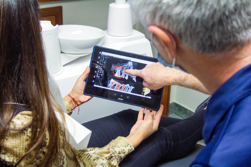
Dentist Glendale, Dr. Sahakyan uses digital dental radiographs to better identify, diagnose, treat and monitor the condition of the oral cavity and disease.
X-Rays are one of the most important diagnostic methods in dentistry.
X-Ray examination, in particular, a panoramic snapshot of the teeth, allows your dentist to see the condition of the teeth, their roots, the way they are located in the bone, and the condition of the bone tissue. Also, the x-ray of the tooth allows you to note various changes in the periodontium, to detect an abscess and a number of other formations, such as cysts and tumors.
Digital X-Ray system, uses less radiation than the majority of imaging provided by other methods.
Why Do You Need Digital X-Rays of Teeth?
- To see at an early stage hidden carious cavities and begin treatment before the tooth becomes sick and begins to harass its master.
- To control the accuracy of the manipulations performed by the doctor. For example, the treatment of the canals, is inconceivable without an X-ray, since only in the teeth picture one can see how the doctor sealed the canals.
- To detect problems with the rudiments of teeth. For example, their incorrect position in the jaw, which in the future can lead to the fact that these teeth cannot erupt in the oral cavity.
- To detect the wrong position of the wisdom tooth. The wisdom tooth is often the cause of the development of crowding of teeth and destruction of the next seventh tooth.
More Detailed Uses of Teeth X-Rays
- Diagnosing caries on the approximal (between the teeth), and subgingival surfaces.
- Diagnostics of secondary caries underseals and crowns.
- Determination of the depth of the carious defect and its relationship with the cavity of the tooth.
- Control of endodontic activities(dental treatment channels).
- Assessment of the condition of periapical (surrounding tooth) bone tissue for periodontal disease and periodontitis.
- Tooth extraction, implantation, prosthetics.
In What Ways is Digital X-Ray Performed?
Digital X-Ray images are obtained in three ways: direct, indirect, and semi-indirect.
The direct method uses an electronic sensor, which is placed in the mouth to create images. The indirect technology uses an X-ray film projector to view traditional dental X-rays as digital images. The semi-indirect digital technology combines a sensor and a scanner, through which X-ray images of teeth are converted into a digital film.
Digital radiographs of the teeth can be made inside (intraoral) or outside (extraoral) the mouth.
Intraoral X-Rays are the most common X-Ray images of teeth. They are used to detect cavities, check the status of teeth development, and monitor teeth and bone state. However, extraoral X-rays do not provide the level of detail as intraoral images provide. Extraoral X-Rays are not used to determine the problems of individual teeth. They are used to determine the retention of the teeth, to track the development and growth of the jaw, and also to find potential problems that arise between the teeth, jaws, and other bones of the face.
Types of Digital X-Rays
In the picture, the doctor can see the location of the implant and uncut teeth, as well as carious cavities in invisible places. The following popular types of digital X-Rays are used by dentist Glendale, at Smile Makeover of LA to diagnose and treat patients:
Bitewing X-Rays
Patients are given bite pictures. For this, the patient needs to bite the plastic X-ray film. In bite pictures, details of the upper and lower teeth in one section of the mouth can be seen. Each bite shows the tooth from the tip to about the level of the supporting bone.
Bitewing intraoral X-Rays are used to:
- determine the destruction between the teeth,
- changes in bone density due to gum disease,
- determine the fit of dental crowns and restorative materials,
- determine the ultimate integrity of the seals.
Periapical X-Rays
Periapical intraoral X-Rays show dentist Glendale, Dr. Sahakyan, the whole tooth. The image starts from the crown to the site where the tip of the root is attached to the supporting bone in the area of the upper or lower jaw. Periapical X-rays are used to determine the structure of the root of the tooth and the abnormality of the surrounding bone structure. Featuring osteoporosis around each tooth, periapical X-Rays are aimed at such conditions as periodontitis, abscesses, and the definition of endodontic metastases.
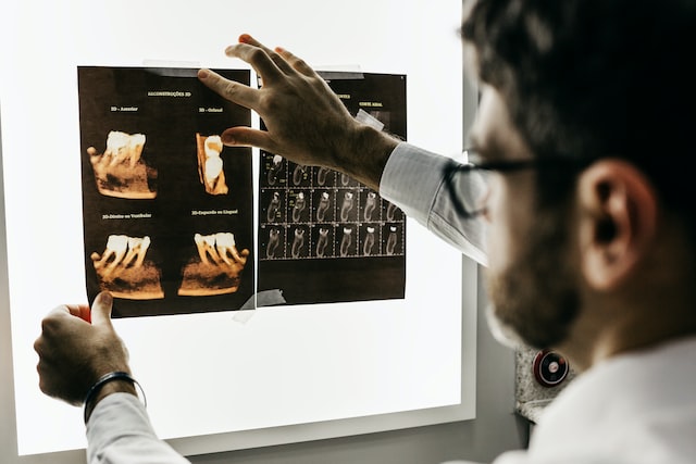
Panoramic X-Rays
Panoramic X-rays are done with a machine that revolves around the head, showing the whole mouth. Panoramic X-Ray, allows the dentist to see not only the teeth but also, the upper jaw joint, the paranasal sinuses, etc. All that is the object of attention of a dentist in Glendale, Dr. Sahakyan at Smile Makeover of LA. Panoramic X-rays are used to design treatment with dental implants, determine retinas of wisdom teeth and jaw problems, and also to diagnose bone and cyst tumors. A panoramic photograph of the teeth is used to solve judicial and legal problems in order to determine bodies that are not recognized in any other way after fires, accidents, and other disasters.
Advantages of Digital X-Rays
- The whole process is much faster than before.
- The preparation of digital images takes much less time than traditional methods.
- Digital X-ray provides a clear and reliable diagnosis and makes the diagnosis more visible and understandable for patients.
- Digital X-rays reveal small hidden areas of destruction between the teeth or under existing seals, bone infections, periodontitis, abscesses or cysts, developmental abnormalities, and tumors that can not only be determined by visual inspection.
- Premature detection and treatment of dental problems can save time, and money and eliminate discomfort.
- The results can be stored in the computer memory, used for comparison at a later time, and rewritten on any media.
- Digital radiographs of teeth are more environmentally friendly than traditional X-Rays, as there is no need to use developers and other chemicals.
- Digital sensors and photostimulating phosphor plates are more sensitive to X-ray radiation and require 60 to 80 percent less radiation than film. This technology holds the principle of achieving results with the minimum possible impact that maintains radiation safety.
- With the help of digital X-ray machines, it is possible to examine both individual teeth and entire parts of the jaw.
Safety
Although with the use of digital radiographs, exposure to radiation is low, no one should receive more radiation than is absolutely necessary. Protective lead aprons and thyroid collars are used to protect the patients from extra radiation.
Smile Makeover of LA is proud to employ the best Dentists and the latest modern dental appliances. Call 1-818-578-2324 to schedule an appointment with a dentist in Glendale and determine the well-being of your teeth.